
Fascia: The Pericardium-Diaphragm Connection
Understanding how the pericardium relates to breathing can in turn help clients relax into their massage session.
The occipitoatlantal (OA) joint is the uppermost weight-bearing synovial joint in the body and is the final junction for adapting to asymmetry or dysfunction from below. Mechanically, the head teeters on the two condyloid joints (shaped like cupped palms tipped slightly medially) that make up the OA joint. This design allows the head to nod back and forth and side to side.
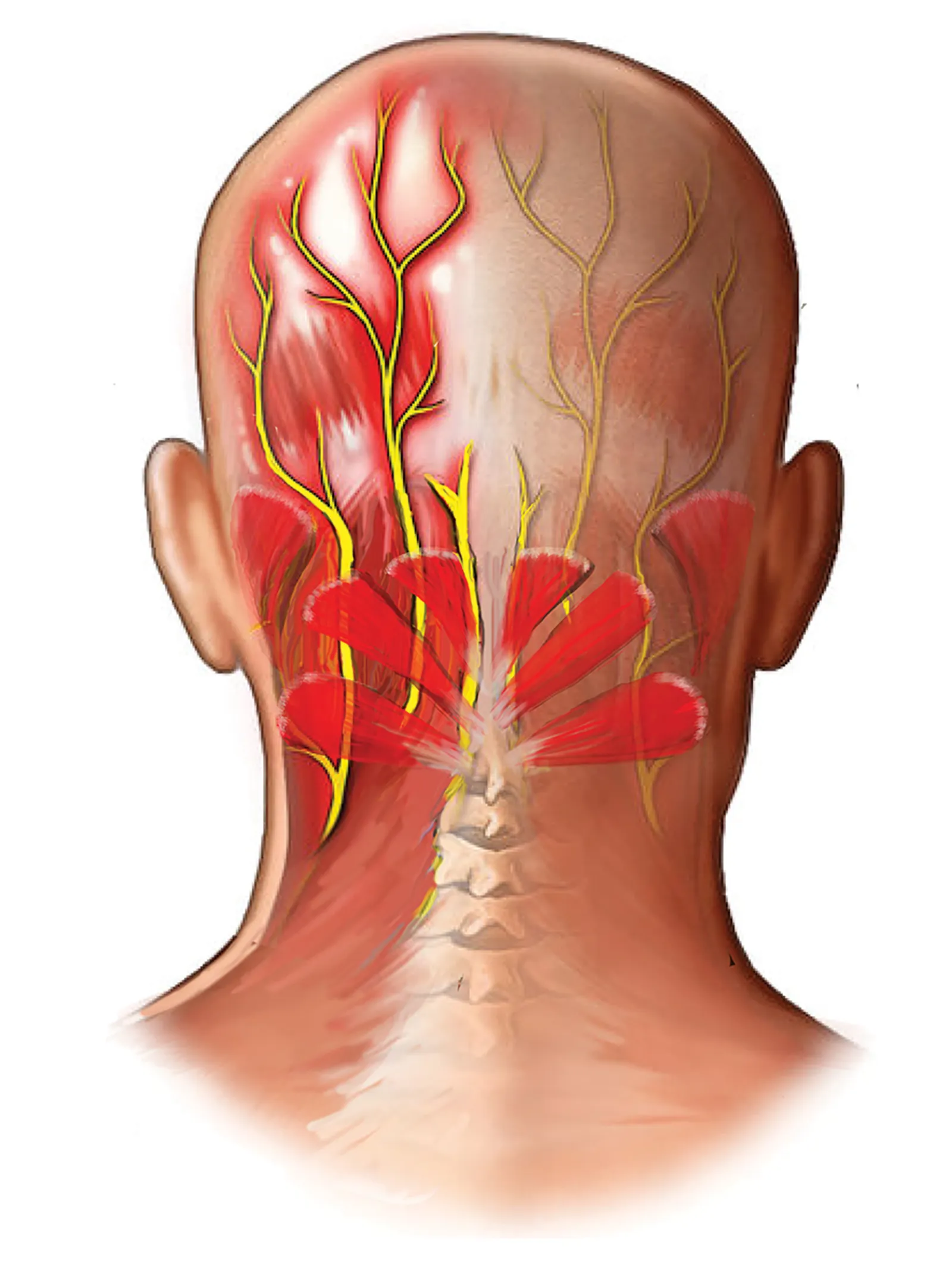
Since the OA joint surrounds and attaches to the brain stem, which controls all basic body functions (including breathing and heartbeat), poor alignment has widespread consequences. For example, if the OA joint on one side develops a restriction, the crooked occiput may set off local neurological reflexes that increase muscle tension in the suboccipitals, and which may compress and irritate the greater occipital nerve. Clients presenting with occipital neuralgia commonly complain of pain referring up the back of the head and around the side, sometimes reaching into the orbital region.
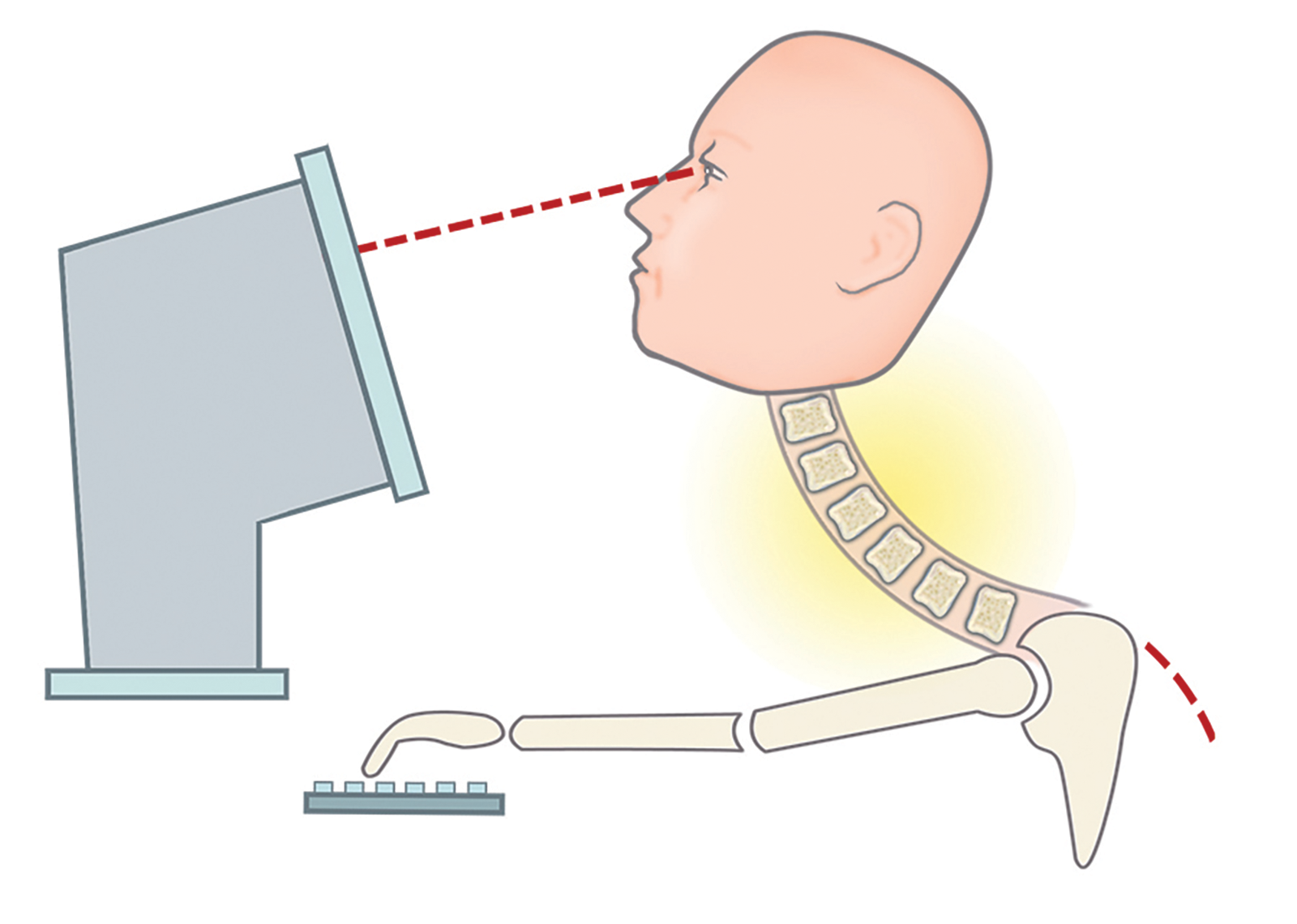
These headaches usually result from holding an awkward, static head posture. Prolonged, non-neutral postures with the head tilted on the neck can cause mechanoreceptor excitability in surrounding muscles, ligaments, and joint capsules as the OA condyles are crammed closed on one side. Examples might include a plumber who works under a sink with his head cocked to one side or an office worker who secures a telephone with her shoulder and chin. Clients whose computer monitors sit either too high or too low may develop bilateral OA restrictions leading to global head pain.
As these activities are unlikely to cause immediate discomfort, clients rarely attribute them as the source of their pain. But palpation by trained hands can tell another story. Suboccipital spasm, tenderness, nodular swellings, and a closing of the inferior nuchal ridge on the posterior arch of the atlas should alert the therapist to test for possible OA jamming.
Before jumping in with knuckles and “fascia-mashing” the suboccipitals, one critical question must first be asked: is the unlevel OA joint and accompanying suboccipital spasm the result of awkward head and neck positioning, as in the case of the plumber under the sink, or could it be compensation for misalignment elsewhere in the body? One way to help solve this mystery is to perform a Brugger Test.
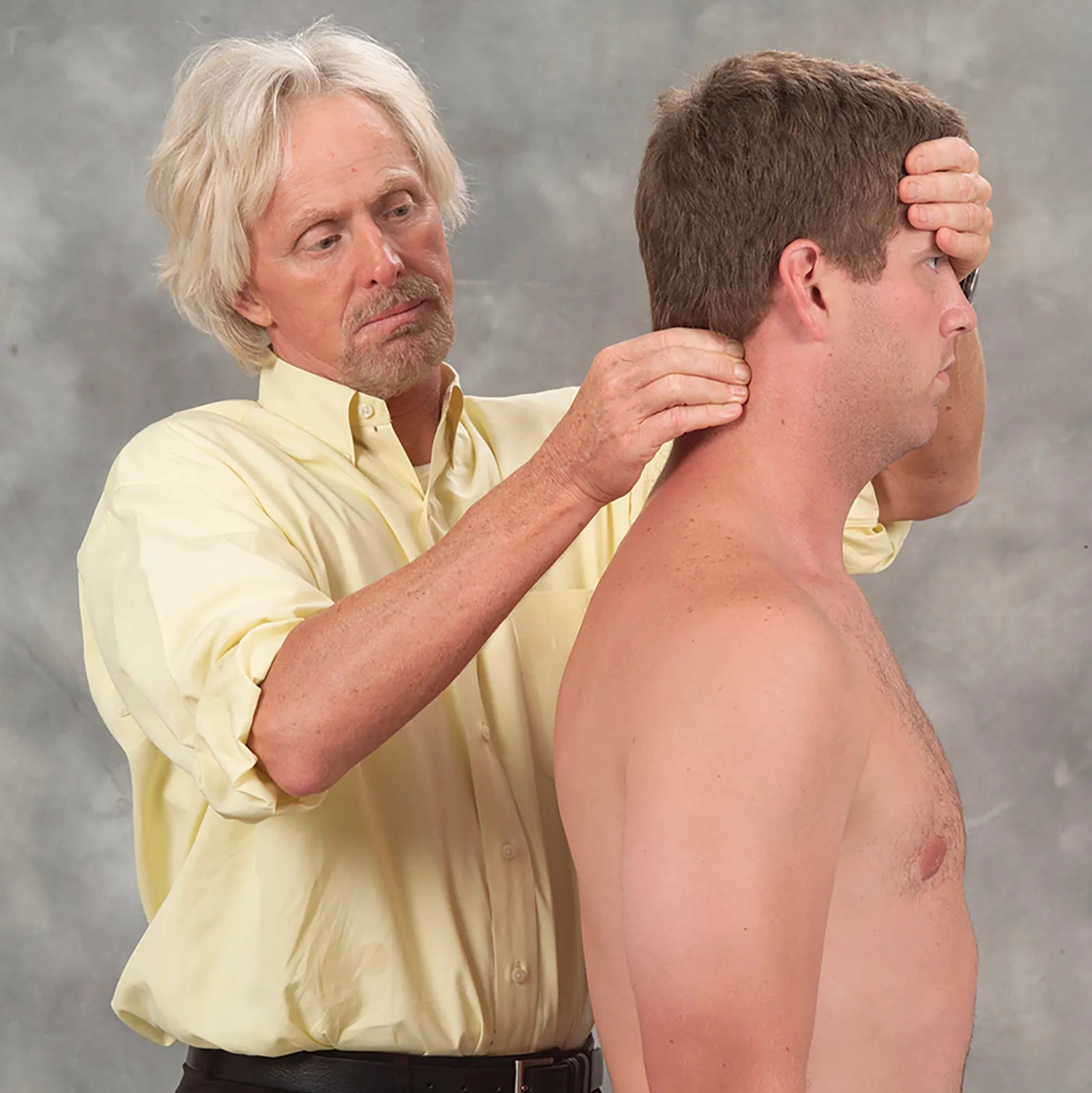
Start with the client standing, with your left hand on the client’s forehead and the index finger and thumb of your right hand gently palpating and holding the knotty suboccipital spasm. Continue to hold this hand position as the client sits. If the spasm decreases when the client sits, this suggests asymmetry somewhere in the lower body is forcing compensation at the OA joint. A functional short leg, unlevel pelvis, or rotoscoliosis are examples of common postural faults that must be addressed before dealing with the fixated OA. If the suboccipital spasm stays knotty when both standing and sitting, it’s an OA problem.
Begin by applying your favorite suboccipital technique to release the protective muscle guarding. Once the suboccipital hypertonicity has calmed, assess and correct OA joint hypomobility by sliding the right hand under the supine client’s head while your left palm secures the forehead. Without lifting the client’s head, ask for a slow chin tuck toward the chest. As the head begins to flex on the neck, follow this movement with your right hand by softly pulling the back of the head superiorly, while the left hand gently pushes the forehead toward the eyebrows—imagine rolling a bowling ball using two hands. By applying a gentle two-second overpressure at the end of this chin tuck maneuver, you should be able to assess the ability of the occipital condyles to glide back on the atlas. If there is restriction to the chin tuck (i.e., a hard restrictive barrier), the OA is unilaterally or bilaterally restricted.
To determine which side is stuck, simply right-sidebend the client’s head 20 degrees and repeat the chin tuck technique. If no resistance is encountered, test the opposite side by left-sidebending 20 degrees and rolling the head into flexion. If the chin does not want to approximate the chest with the head in this position, then the right occipital condyle is stuck anteriorly and is unable to glide back on the right atlas condyle. If the client’s pain is also on the right, the fixated right condyle is likely the main event causing occipital neuralgia.
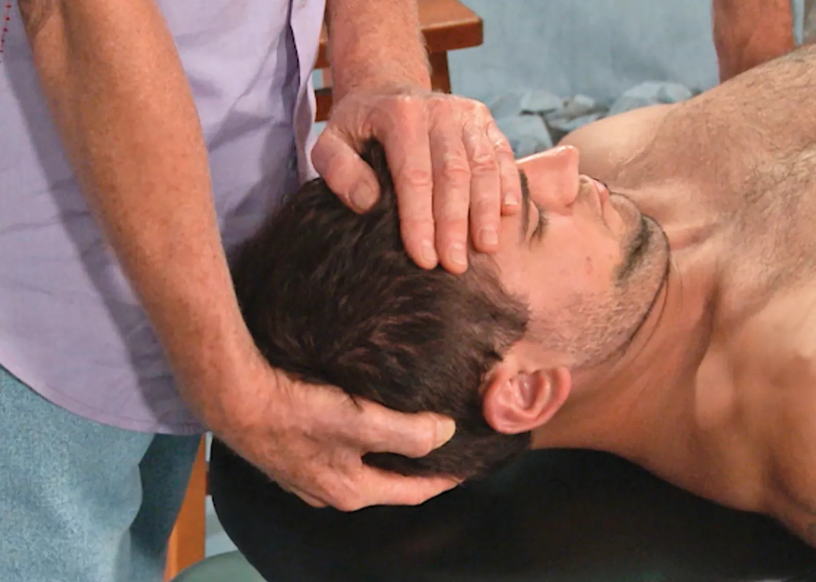
To free the soft tissues that are restraining joint motion, keep the client’s head in the same left-sidebent position and ask for a chin tuck while your hands follow with two seconds of gentle overpressure at the end range of flexion (stop if the client experiences any discomfort). Repeat three times and retest with the head in a neutral position.
Your clients will love this gentle myoskeletal O-A stretch and appreciate relief from the nagging, and sometimes debilitating, pain of occipital neuralgia.
Understanding how the pericardium relates to breathing can in turn help clients relax into their massage session.
Superficial fascia may not get the same attention as deep fascia, but it plays an extremely important role in the organization of tissues and the structure of the body.
Understanding tendons—their shapes, lengths, and organization—improves an MT’s touch vocabulary and facilitates a more skilled touch.
While the neck is a bridge, a pathway, the position of the neck and head can also indicate a multitude of other things happening beneath the surface.