


Fascia: The Pericardium-Diaphragm Connection
Understanding how the pericardium relates to breathing can in turn help clients relax into their massage session.
The teres major is a muscle on the back of the shoulder that joins with the latissimus dorsi to form the posterior axillary fold. Compared to the latissimus dorsi, the teres major is much smaller and spans less distance. It has a specific origin on the inferior portion of the lateral border of the scapula, while the latissimus dorsi has a broad attachment on the trunk. This origin is just inferior to that of the teres minor, with the long head of the triceps brachii running between the two teres muscles. Both the latissimus dorsi and the teres major wrap around the humerus anteriorly and insert on the lesser tubercle and bicipital groove, with the teres major inserting slightly more posteriorly.
Though they differ in size and shape, the teres major and the latissimus dorsi share all the same actions: extension, adduction, and internal rotation of the shoulder. The teres major is often called “lat’s little helper” because of the strong functional association. It is a prime mover muscle and is most active when pulling the arm down and back from an overhead position or adducting the arm directly into the body when the scapula is fixed.
Although the teres minor is located near the teres major, and has a similar shape, these muscles do not share functions.
When the arm is fixed, the teres major and latissimus dorsi work together to pull the trunk toward the arm—for example, in pull-ups and climbing. When the arm is free, the teres major works with other shoulder internal rotators and extenders (the lower fibers of the latissimus dorsi, pectoralis major, and subscapularis) to pull the raised arm forward and downward—for example, in swimming, hitting, or throwing overhead.
The teres major is particularly active when maintaining position of the upper extremity. This static activation is common in computer work, driving, or other activities where the arm is held steady in a given position. Fatigue from prolonged positioning may lead to adhesions, chronic pain, hypertonicity, ischemia, and trigger points in the posterior shoulder and particularly the teres major. The area of muscle convergence in the posterior axillary fold is commonly tender and ischemic, and contains numerous trigger points.
Targeted manipulation and active movement (such as the cat/cow pose) can help flush the tissue, decrease pain, and restore function.
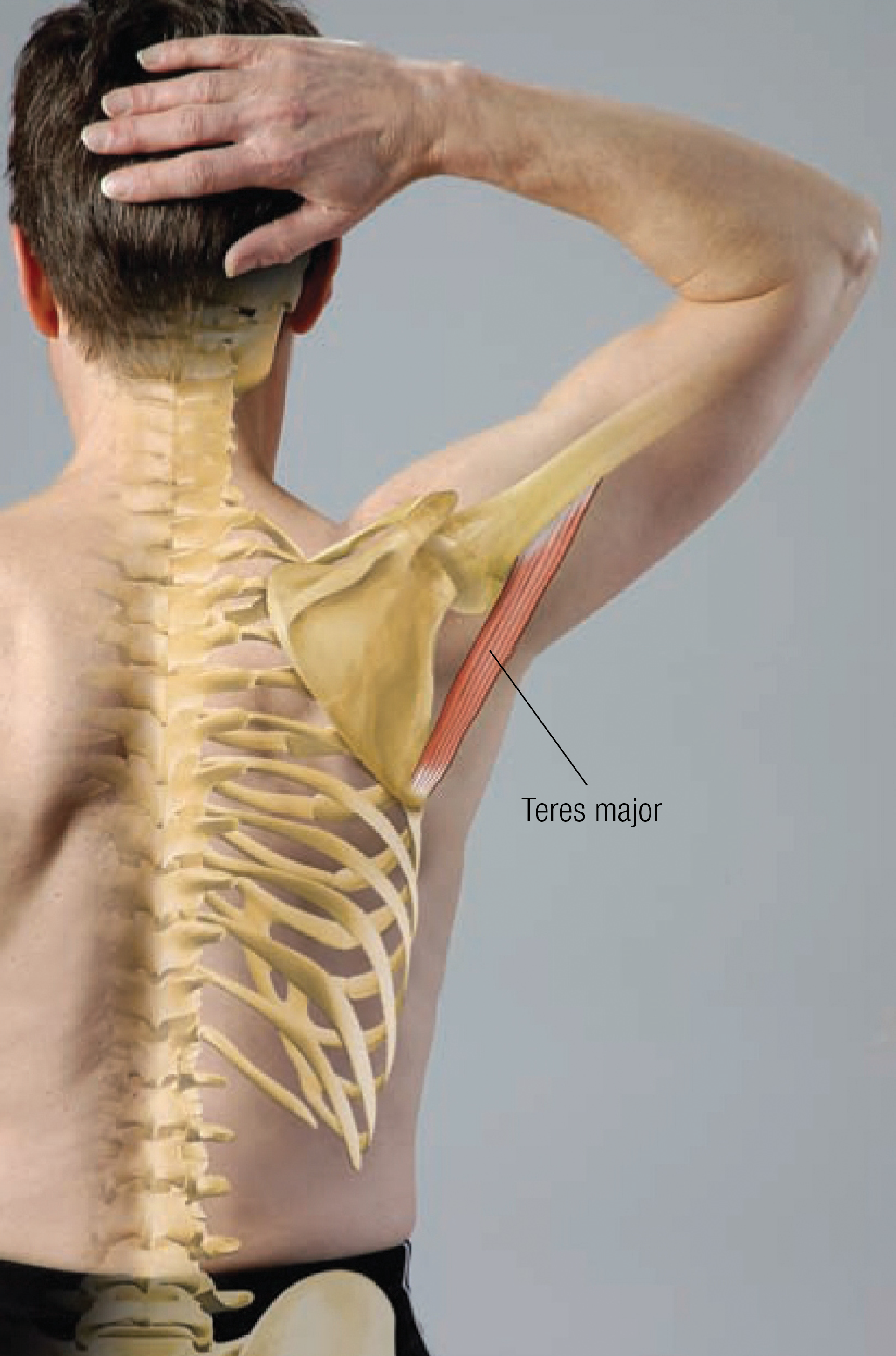
Attachments
Actions
Innervations
Positioning: client prone with arm at side.

Editor’s note: The Client Homework element in Functional Anatomy is intended as a take-home resource for clients experiencing issues with the profiled muscle. The stretches identified in Functional Anatomy should not be performed within massage sessions or progressed by massage therapists, in order to comply with state laws and maintain scope of practice.
Understanding how the pericardium relates to breathing can in turn help clients relax into their massage session.
Superficial fascia may not get the same attention as deep fascia, but it plays an extremely important role in the organization of tissues and the structure of the body.
Understanding tendons—their shapes, lengths, and organization—improves an MT’s touch vocabulary and facilitates a more skilled touch.
While the neck is a bridge, a pathway, the position of the neck and head can also indicate a multitude of other things happening beneath the surface.