

Exploring Tendons
Understanding tendons—their shapes, lengths, and organization—improves an MT’s touch vocabulary and facilitates a more skilled touch.
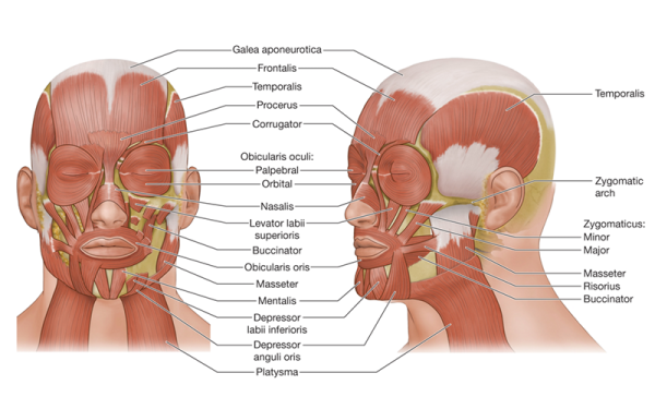
The facial muscles lie just under the skin and cover the anterior and lateral skull and mandible. This complex network of muscles performs a variety of functions, including moving the jaw, mouth, nostrils, eyelids, and scalp. They also create a wide array of facial expressions—raising your eyebrows, squinting your eyes, pursing your lips, and even wiggling your ears—and are subject to the same issues that affect other muscles, like hypertonicity, adhesions, and trigger points.
In addition to the direct actions generated by the facial muscles, there is a well-established functional connection between the temporomandibular and craniocervical regions of the body.1 This means that because facial muscles create movement and direct the positioning of the temporomandibular joint (TMJ), they also influence posture and movements in the head and neck. Preprogrammed neurological commands coordinate movements of the jaw, head, and neck, making the health and proper function of one dependent on that of the others. Traumatic injuries and chronic conditions affecting one of these areas will also directly affect the others.
Beyond the interconnectedness of the face, head, and neck, there are other unique neurological features of the facial region. When examining how much area within the brain is used to process sensory input and direct motor control of different regions of the body, the space dedicated to the face is relatively large. Both the hands and the face have high sensory importance, which is identified by how richly the skin is innervated. Wilder Penfield, MD, developed a three-dimensional map of the disproportionate distribution of sensory importance in a figure identified as a cortical homunculus.
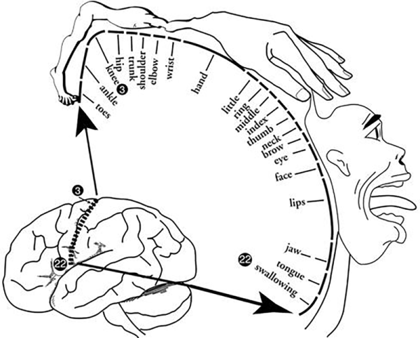
Observing this figure, there are implications as to the way we approach bodywork and specifically how we organize our time during a session. Clients often come in wanting extra attention on their neck or back, or even their feet. Since we are often constrained by a set session time, other areas will receive less attention as we honor the client's wishes. If we consider the sensory importance of different areas, which areas are most likely to have the biggest neurological influence? Is this consistent with how we approach timing full-body treatments? What about our approach to treating TMJ dysfunction or whiplash?
Based on the constant utilization and complexity of movements activated by the facial muscles, the sensory importance of the facial region, and the strong connection between the facial muscles and the health and function of the head, neck and jaw, it may be worthwhile to revisit the amount of time spent working on this area.
Specifically, focused work on the face and scalp can enhance parasympathetic activity and also improve efficacy of treatments for both traumatic injuries and chronic conditions affecting the head, neck, and jaw.
1. H. Zafar, "Integrated Jaw and Neck Function in Man. Studies of Mandibular and Head-Neck Movements During Jaw Opening-Closing Tasks," Swedish Dental Journal 143 (Supplement) (2000): 1-41.
Understanding tendons—their shapes, lengths, and organization—improves an MT’s touch vocabulary and facilitates a more skilled touch.
While the neck is a bridge, a pathway, the position of the neck and head can also indicate a multitude of other things happening beneath the surface.
Understanding fibroblasts and the extracellular matrix changes how we think about the tissue we touch.
Studies reveal that 37 percent of the force generated by muscle contraction is transmitted to adjacent connective tissue structures instead of the bones.